Arteries Diagram Upper Body / Arteries Of The Upper Limb The Lecturio Medical Online Library - Left main coronary artery branches into the circumflex artery and the left anterior descending artery.
byAdmin•
0
Arteries Diagram Upper Body / Arteries Of The Upper Limb The Lecturio Medical Online Library - Left main coronary artery branches into the circumflex artery and the left anterior descending artery.. Subclavian artery the subclavian artery is the large vessel that begins the blood supply to the upper extremity. An artery (plural arteries) (from greek ἀρτηρία (artēria) 'windpipe, artery') is a blood vessel that takes blood away from the heart to one or more parts of the body (tissues, lungs, brain etc.). It begins near the heart and travels under the clavicle bone toward the shoulder. Your body organs range from your brain, heart, liver, skin, lungs, kidneys, intestines, stomach, bladder, etc. The human torso is also known as the 'trunk'.
The brachial artery is a continuation of the axillary artery past the lower border of the teres major. Left main coronary artery branches into the circumflex artery and the left anterior descending artery. The video demonstrates first veins and then arteries of the upper thorax and right upper limb. The human torso is also known as the 'trunk'. Some of the most critical human body organs are situated within the torso.
Chap 18 Blood Vessels Continued Learning Objectives Continued 1 Name And Give The Location Of The Major Arteries And Veins In The Systemic Circulation Ppt Download from images.slideplayer.com Some of the most critical human body organs are situated within the torso. After removing the flexor digitorum superficialis, we clearly see the ulnar artery and nerve, median nerve and radial artery. Smaller arteries are more muscular in the structure of their walls. These are protected by the rib cage. The human torso is also known as the 'trunk'. The heart is responsible for pumping blood throughout the body using a complex system of veins and arteries. As the heart pumps inside the center of the chest, oxygenated blood circulates to organs and other tissues starting with the aorta, the largest artery in the body, and arteries that branch from it. Left main coronary artery branches into the circumflex artery and the left anterior descending artery.
The two exceptions are the pulmonary and the umbilical arteries, which carry deoxygenated blood to the organs that oxygenate it (lungs and placenta.
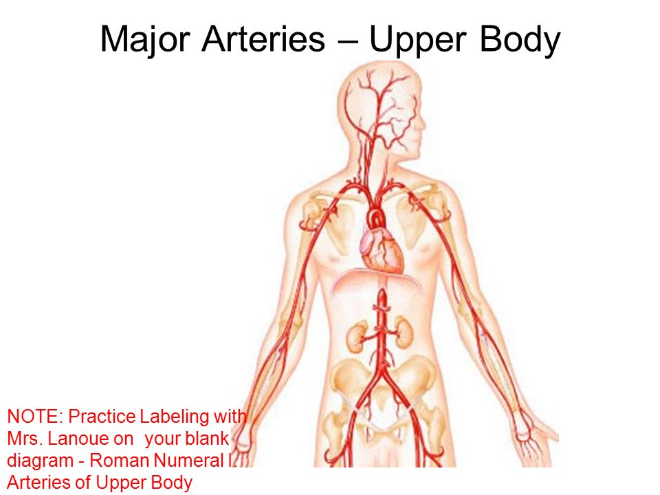
The aorta is the largest artery in the body and is divided into 3 parts: This diagrams shows the major arteries in the human body. Arteries serving the upper limbs; Some of the most critical human body organs are situated within the torso. The stomach is found on the left side of the body in the upper abdomen region. The human torso is also known as the 'trunk'. Arteries & veins of the upper body 1. See more ideas about anatomy and physiology, medical anatomy, medical knowledge. Most arteries carry oxygenated blood; The brachial artery is a continuation of the axillary artery past the lower border of the teres major. It begins near the heart and travels under the clavicle bone toward the shoulder. Continuation of the subclavian artery as it penetrates the body wall and enters the axillary region; An artery (plural arteries) (from greek ἀρτηρία (artēria) 'windpipe, artery') is a blood vessel that takes blood away from the heart to one or more parts of the body (tissues, lungs, brain etc.).
Immediately distal to the teres major, the brachial artery gives rise to the profunda brachii (deep artery), which travels with the radial nerve in the radial groove of the humerus and supplies structures in the posterior. Inferior vena cava vein carrying blood deoxygenated in the lower portion of the body (below the diaphragm) to the right atrium; The human torso is also known as the 'trunk'. See more ideas about anatomy and physiology, medical anatomy, medical knowledge. Arteries carry blood away from the heart in two distinct pathways:
Circulatory System Arteries Of The Upper Limb Flow Chart Youtube from i.ytimg.com The heart is responsible for pumping blood throughout the body using a complex system of veins and arteries. Inferior vena cava vein carrying blood deoxygenated in the lower portion of the body (below the diaphragm) to the right atrium; Continuation of the subclavian artery as it penetrates the body wall and enters the axillary region; The heart has four different chambers. See more ideas about anatomy and physiology, medical anatomy, medical knowledge. Second segment of the aorta, which branches into the arteries flowing to the head and upper limbs; This diagram of the human heart shows all the major vessels, and arrows indicate the direction of flow through the heart. The two upper chambers are called atria, and the two lower chambers are called ventricles.
It begins near the heart and travels under the clavicle bone toward the shoulder.
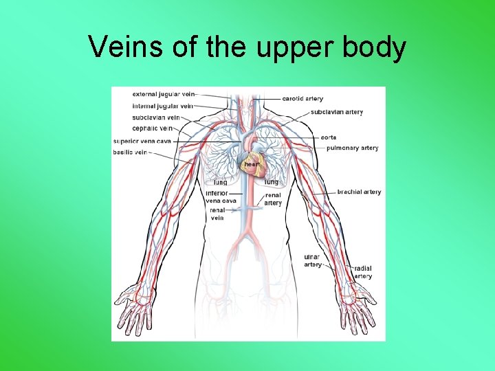
Main branches from the aorta include the brachiocephalic artery, left carotid artery, and the left subclavian artery. The video demonstrates first veins and then arteries of the upper thorax and right upper limb. Arteries & veins of the upper body 1. Veins and arteries in the arm diagram veins in the arm new brachial artery and anastomoses around elbow arteries and veins of the arm smartdraw create healthcare diagrams like this example. The stomach is found on the left side of the body in the upper abdomen region. The two upper chambers are called atria, and the two lower chambers are called ventricles. The hand anatomy enables us various movements. The brachial artery is a continuation of the axillary artery past the lower border of the teres major. The aorta is the largest artery in the body that exits the left ventricle of the heart. The aorta is the largest artery in the body and is divided into 3 parts: The smooth muscles of the arterial walls of these smaller arteries contract or expand to regulate the flow of blood through their lumen. Some of the most critical human body organs are situated within the torso. Ninja nerds,join us in this video where we discuss the arteries of the upper limb in a flow chart style.
The brachial artery is a continuation of the axillary artery past the lower border of the teres major. The arteries of the upper extremity. Test your knowedge of the arteries of the upper limb with the labeling page. The majority of the vessel continues into the brachium and becomes the. Left main coronary artery branches into the circumflex artery and the left anterior descending artery.
Cardiovascular System Cardiac Output And Everything Else Dr from slidetodoc.com This diagrams shows the major arteries in the human body. 5 draw labelled diagram to show the arterial supply of internal capsule. The chapter on the upper limb arteries begins with an overview of the arteries (subclavian, axillary, brachial, radial and ulnar arteries and deep and an anatomical diagram illustrates the lymph nodes of the upper limb, with the pectoral, axillary (lymphatic ganglia), paramammary, parasternal, humeral. Your body organs range from your brain, heart, liver, skin, lungs, kidneys, intestines, stomach, bladder, etc. The brachial artery is a continuation of the axillary artery past the lower border of the teres major. Ninja nerds,join us in this video where we discuss the arteries of the upper limb in a flow chart style. The heart has four different chambers. Left main coronary artery branches into the circumflex artery and the left anterior descending artery.
The video demonstrates first veins and then arteries of the upper thorax and right upper limb.
The smooth muscles of the arterial walls of these smaller arteries contract or expand to regulate the flow of blood through their lumen. The hand is probably the finest product of human evolution from the aspect of our body mechanics. The ascending aorta, arch of the aorta, and descending aorta. Smaller arteries are more muscular in the structure of their walls. In this image, you will find external carotid artery, internal carotid artery, vertebral artery, aorta and arch, pulmonary artery, cardiac artery, thoracic aorta, celiac trunk, superior mesenteric artery, renal artery, gonadal artery, inferior mesenteric artery, common iliac artery, external iliac artery, internal iliac artery, deep femoral artery, femoral artery, popliteal artery, anterior tibial artery, peroneal artery, posterior tibial artery. Left main coronary artery branches into the circumflex artery and the left anterior descending artery. The two exceptions are the pulmonary and the umbilical arteries, which carry deoxygenated blood to the organs that oxygenate it (lungs and placenta. Veins and arteries in the arm diagram veins in the arm new brachial artery and anastomoses around elbow arteries and veins of the arm smartdraw create healthcare diagrams like this example. This diagrams shows the major arteries in the human body. Arteries & veins of the upper body 1. The brachial artery is a continuation of the axillary artery past the lower border of the teres major. The aorta is the largest artery in the body that exits the left ventricle of the heart. Aortic arch<br />what are the arteries that come off the aortic arch in order?<br />brachiocephalic trunk<br />turns into the right subclavian and right common carotid arteries.<br />left common carotid artery <br />left subclavian artery<br />
The ascending aorta, arch of the aorta, and descending aorta arteries diagram. Test your knowedge of the arteries of the upper limb with the labeling page.